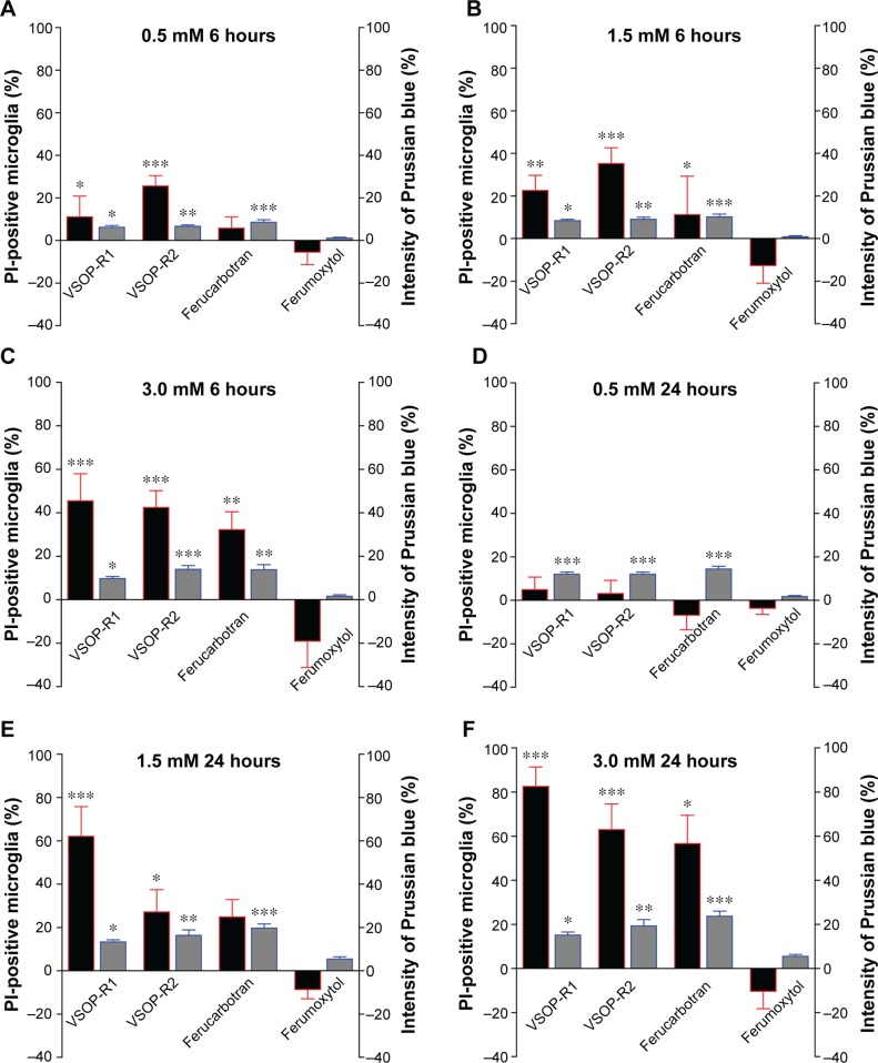
Figure 2.
The viability of primary microglia was SPIO-dependent.
Notes: Microglia were incubated for either 6 hours (A–C) or 24 hours (D–F) with SPIOs of various concentrations. Black red-framed bars indicate the percentage of PI-positive, nonviable cells (left y-axis in each plot); gray blue-framed bars indicate the percentage of Prussian blue staining intensity, referring to microglial iron content (right y-axis). All values are normalized to untreated microglia. (A–F) Iron accumulation of microglia exposed to VSOPs and ferucarbotran significantly increased compared to microglia exposed to ferumoxytol after 6 or 24 hours. Extended incubation from 6 to 24 hours led to an increase in the number of PI-positive cells by more than 20% for all SPIOs except ferumoxytol. Note that increases in SPIO accumulation do not correspond with proportional increases in the numbers of nonviable microglia. Kruskal–Wallis one-way analysis of variance and Dunn’s multiple comparison post hoc test, expressed as mean ± standard error of mean: (A–F) ***P<0.0004; **P<0.01; *P<0.05.
Abbreviations: SPIO, superparamagnetic iron oxide nanoparticle; PI, propidium iodide; VSOPs, very small iron oxide particles.

