Figure 3.
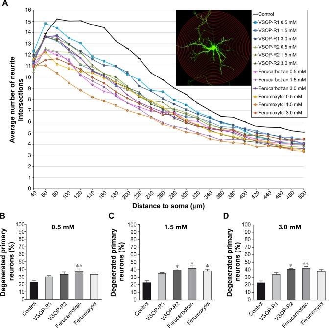
Primary neurons degenerated after SPIO exposure.
Notes: (A) Sholl analysis of primary neurons reveals reduced numbers of neuronal processes after SPIO exposure for 24 hours. Neurite intersections per exposure condition were counted manually using 24 20 μm-spaced concentric circles, as shown in the immunofluorescence example image of a neuron. (B–D) SPIO- and concentration-specific decreases in neuronal viability were observed after quantification of the percentage of degenerated neurons with fluorescence microscopy. This effect was independent of the SPIO type. Kruskal–Wallis one-way analysis of variance and Dunn’s multiple comparison post hoc test, expressed as means ± standard error of mean: (B) **P#0.0174; (C) *P<0.05; (D) *P<0.05, **P#0.0029.
Abbreviations: SPIO, superparamagnetic iron oxide nanoparticle; VSOP, very small iron oxide particle.

