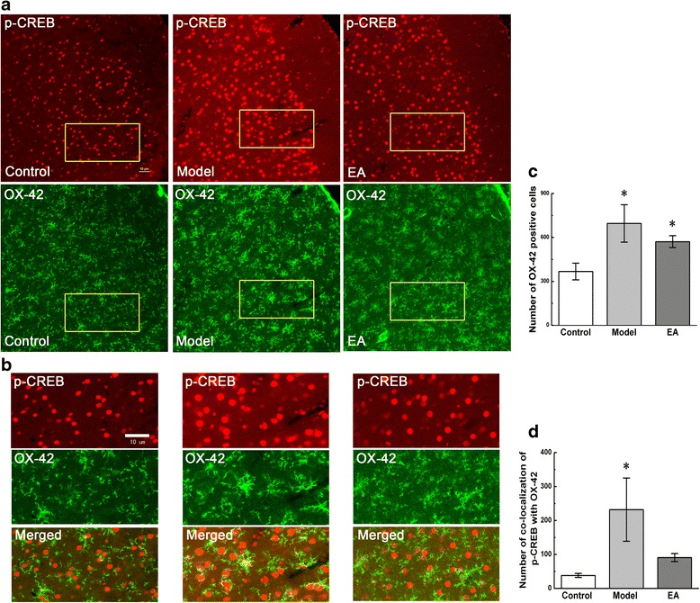
Figure 5.

Co-localization of p-CREB and OX-42 in coronal brain sections of ACC. Photomicrographs showed the expression of p-CREB (red) and OX-42 (a microglial marker, green) from the same sections in figure a. Figure b was a high magnification image of the areas indicated by the yellow squares in the figure a. The double-immunofluorescence labeling showed that p-CREB co-expressed with OX-42 in the ACC. Numbers of OX-42-positive cells and co-localization of p-CREB with OX-42 were analysed in figure c and d. Error bars indicated standard error of the mean. Four rats for each group, five slides for each rat. * p < 0.05 vs. the control group.
