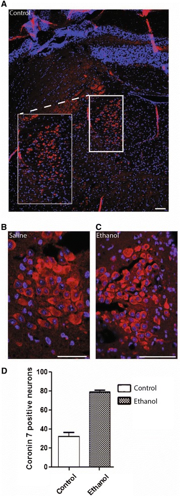
Figure 3.

Expression of Coro7 in locus coeruleus (LC) in mouse treated with ethanol. Mice were treated with saline or ethanol and cells expressing Coro7 in the locus coeruleus was visualized using Coro7 antibody. (A) Brain was cut coronal and the locus coeruleus is shown as overview. (B) close up from saline treated and (C) ethanol-treated mice. (D) The number of positive cells were counted in LC and displayed as a column chart. Error bars indicate SD. Significance levels are indicated: *P < 0.05; **P < 0.01; ***P <0.001. Each group contained eight male mice. Scale bar: 0.1 mm.
