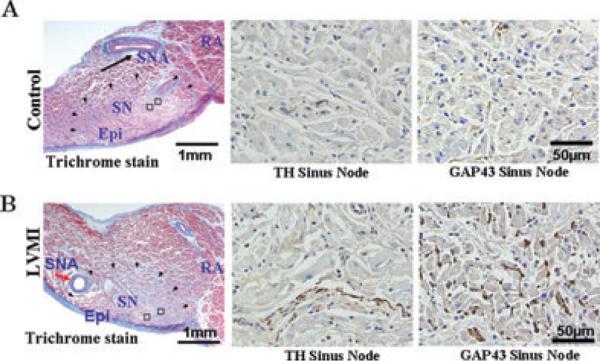
Figure 6.
Tyrosine hydroxylase (TH) and growth associated protein43 (GAP43)-positive staining nerves. Panels A and B are in the sinus node region and panels C and D are in the LSPV and the LAA. The open squares indicate the regions of immuno-stained histological sections shown to the right of each panel. Notice the considerable increase in both TH and GAP43 positive nerves in atria isolated from dogs with chronic LVMI compared to atria isolated from sham-operated dogs at all 3 sites, sinus node (panel A and B), the LSVP and the LAA (panels C and D). Epi epicardium; RA right atrium; SN = sinus node; SNA = sinus node artery.

