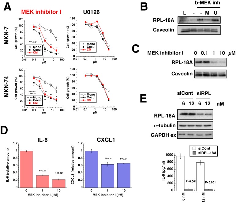
Fig 3. Effect of MEK inhibitor I on co-culture of gastric cancer cells with gastric stromal cells.
(A) MKN-7 and MKN-74 cells were cultured alone (Mono) or co-cultured with Hs738 (Cocul) for 3 days in the presence of MEK inhibitor I or U0126. Hs738 cells were cultured with the inhibitors for 2 days and CM was prepared. Both gastric cancer lines were cultured in the CM for 3 days. Cell growth was determined by measuring GFP fluorescence intensity. The values are means ± s.d. (n = 3). Cell growth is expressed as a percentage of the value without test compounds in each culture condition. (B) Hs738 cell extracts were incubated with b-MEK inh-pretreated streptavidin resin in the presence or absence of MEK inhibitor I (M) or U0126 (U) and the bound proteins were analyzed by Western blotting. L, 1/50 of loaded cell extracts. (C) Hs738 cells were cultured with MEK inhibitor I for 2 days and the cell lysates were analyzed by Western blotting. (D) Hs738 cells were cultured with MEK inhibitor I for 2 days and the concentrations of IL-6 and CXCL1 in the CM were determined. The values are means ± s.d. (n = 3). (E) Hs738 cells were treated with siRNA specific for RPL-18A (siRPL) or negative control (siCont) for 2 days and then re-inoculated followed by further culture for 2 days. The cell lysates were analyzed by Western blotting and the amounts of IL-6 in the CM were determined. The values are means ± s.d. (n = 3).

