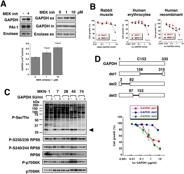
Fig 6. Gastric stromal cells secreted GAPDH and suppressed the growth of gastric cancer cells.
(A) Concentrated Hs738 CM prepared by culturing with or without 10 μM MEK inhibitor I was analyzed by Western blot (upper left). Hs738 cells were cultured with MEK inhibitor I for 2 days and the cultured supernatant was collected. GAPDH and enolase in the culture supernatant (ex) and the cell lysates (in) was analyzed by Western blot (upper right). GAPDH enzyme activity was examined in the cultured supernatant (lower). The values are means ± s.d. (n = 3). (B) MKN-7 and MKN-74 cells were cultured with various amounts of GAPDH for 3 days. Cell growth was determined using MTT. The values are means ± s.d. (n = 3). (C) Gastric cancer cells were cultured with or without human erythrocyte GAPDH at 5 U/mL for 1 day. The cell lysates were analyzed by Western blotting with anti-phospho-Ser/Thr antibody (9624) and the indicated antibodies. An arrowhead indicates the position of RPS6. (D) MKN-7 cells were cultured with human recombinant deletion mutants of GAPDH for 3 days. Cell growth was determined using MTT. The values are means ± s.d. (n = 3). Cell growth is expressed as a percentage of the value without GAPDH in each culture condition.

