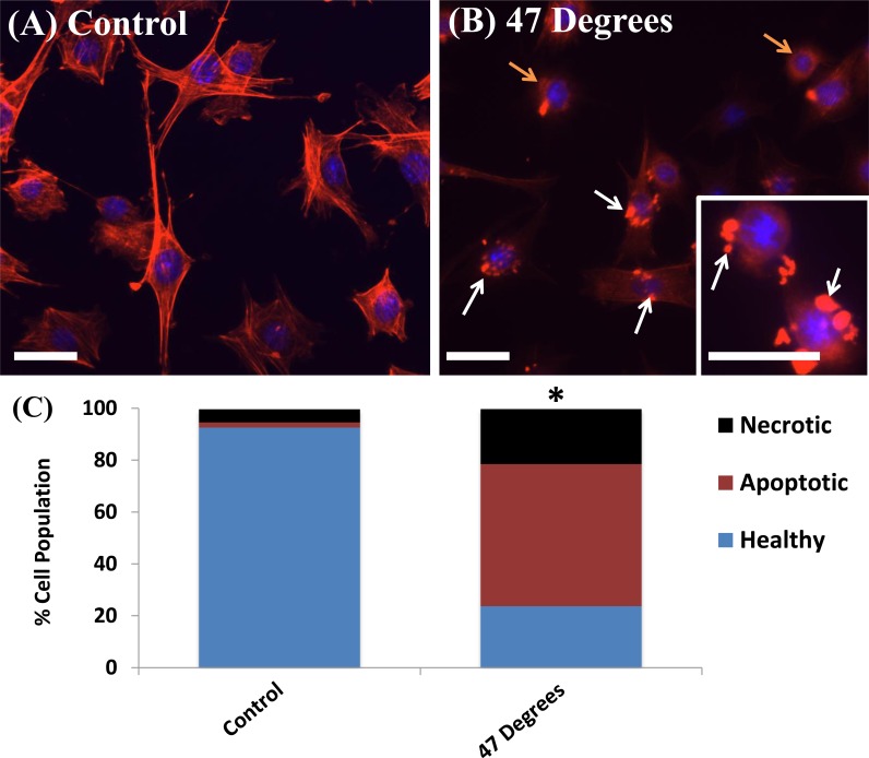
Fig 1. Heat-treatment induces damage responses in MLO-Y4 cells.
Phalloidin stained actin filaments (red) and DAPI stained nucleus (blue) of MLO-Y4 cells heat-treated to (A) 37°C (control) and (B) 47°C for 1 minute demonstrating membrane condensation (white arrows) and rounded cell bodies detaching (orange arrows). Scale bar = 32μm. (C) Flow cytometry quantification of necrotic, apoptotic and healthy cell populations 24 hours after heat-treatment. * indicating statistical difference in the number of viable, apoptotic and necrotic cells compared to the 37°C (control) (p ≤ 0.05).

