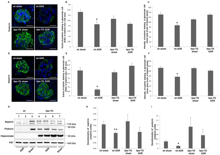
Fig 2. Expression of key slit diaphragm proteins is reduced in wild type but not in hpa-TG Adriamycin-injected mice.
Podocyte markers expression is reduced in wild type but not in hpa-TG Adriamycin-injected mice. wt or hpa-TG mice were injected with Adriamycin (ADR) or served as control (sham). Two weeks post injection, the animals were sacrificed. (A, D) Representative immunofluorescence staining performed on cryostat sections from kidneys of the indicated experimental groups, using anti-podocin or anti-nephrin primary antibodies, and Dylight 488-conjugated anti-rabbit IgG. Nuclei were stained with DAPI (Confocal microscope, scale bar = 25 μm). The extent of podocin (B) and nephrin (E) staining and its intensity (C, F) in glomerular tuft were quantified. For podocin we analyzed n = 30, 23, 15 and 24 glomeruli, for wt sham, wt ADR, hpa-TG sham, and hpa-TG ADR mice, respectively), whereas for nephrin n = 22, 23, 17 and 27 glomeruli, for wt sham, wt ADR, hpa-TG sham, and hpa-TG ADR mice, respectively. Glomeruli were evaluated at least from 3–4 mice per each experimental group. *, P<0.01 vs. wt sham. (G) Protein was extracted from kidney cortex or isolated glomeruli and analyzed by Western blotting using the indicated antibodies. P97 was used as loading control. A representative immunoblot is depicted in panel G (n = 7, 11, 7 and 11 for wt sham, wt ADR, hpa-TG sham, and hpa-TG ADR mice, respectively from three independent experiments) (H, I) Densitometry of immunoblotting of isolated glomeruli lysates (n = 8, 7, 3 and 6 for wt sham, wt ADR, hpa-TG sham, and hpa-TG ADR mice, respectively). *, P = 0.03 vs. wt sham **, P = 0.02 vs. wt sham.

