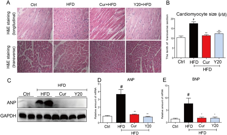
Fig 5. Y20 attenuates cardiac histological abnormalities and hypertrophy in the hearts of HFD-fed rats.
(A) Representative images for the Hematoxylin-Eosin (H&E) staining in the formalin-fixed myocardial tissues (400× magnification). (B) Qantitative data of myocyte cross-section lenth of 100 cells chosen from different visual scopes of 4 samples per group in myocardial transverse H&E staining were shown. (C) Western blot analysis for the protein expression of ANP in the myocardial tissues was performed. (D & E) The mRNA expression of the hypertrophic markers ANP and BNP in the myocardial tissues was detected by real-time qPCR. Four rats in each group were used for above analysis. *, P<0.05, **, P<0.01 v.s. HFD; # P<0.05 v.s. vehicle control (Ctrl).

