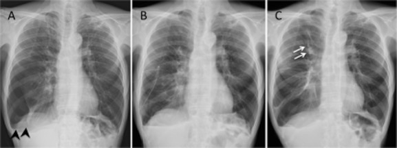
Figure 1.

Chest radiographs for case 1. Chest radiograph on admission showed pneumothorax in the right lower lung field (arrowheads) (A). After talc poudrage, the right lung was re-expanded (B). Chest radiograph taken 1 month after chest tube removal followed by bronchial occlusion with endobronchial Watanabe spigot (EWS) to the right B1a (arrows) showed complete lung re-expansion (C).
