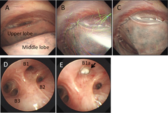
Figure 2.

Endoscopic images for case 1. Pleuroscopy before talc poudrage revealed the right upper lobe was adhered to the chest wall (A). Talc was insufflated pleuroscopically under visualization (B). At the end of the procedure, talc was well distributed on the pleura (C). Bronchoscopic image of the right upper bronchus (D). Right B1a was occluded with endobronchial Watanabe spigot (EWS) (arrow) (E).
