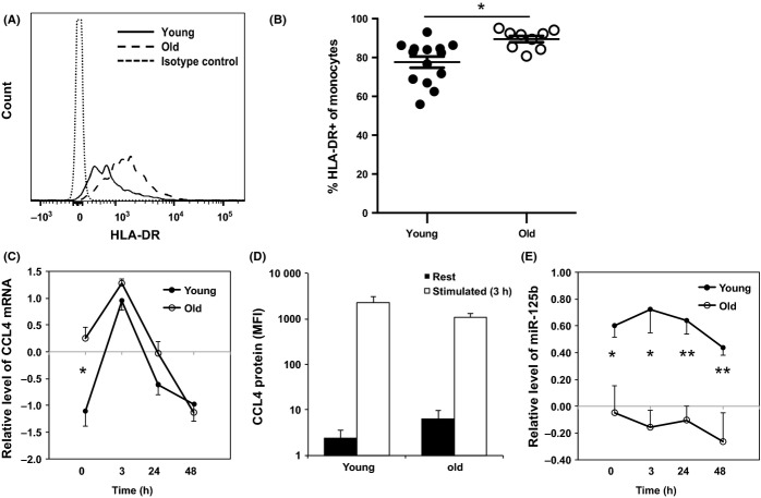
Figure 2.
Activation-induced change of CCL4 and miR-125b in monocytes from young and old adults. (A) A representative histogram of HLA-DR staining on monocytes from young and old adult. The positive staining of HLA-DR was based on the isotype control staining. (B) Percentages of HLA-DR+ monocytes in young (N = 14) and old (N = 9) adults. Whole blood samples were stained with antibodies against CD14, HLA-DR, and other markers, analyzed by FACSCaton II, and percentages of HLA-DR+ in monocytes were presented. (C) Relative levels of CCL4 mRNA in monocytes after in vitro stimulation. The relative levels (in Log10) were after normalization to a standard generated from PBMCs of five normal subjects. (D) Intracellular CCL4 protein staining of freshly isolated (rest) and LPS stimulated (3 h) monocytes. The average mean fluorescent intensity (MFI) is shown (young = 6 and old = 11). (E) Relative levels of miR-125b in monocytes after in vitro stimulation. Freshly isolated monocytes from young (N = 6) and old (N = 6) adults were stimulated with LPS for 3, 24, and 48 h. The levels of CCL4 mRNA and miR-125b were determined by RT-qPCR and normalized to ACOX-1 and to RNU6B first and then normalized to a standard generated from PBMCs of five normal subjects in Log10 scale, respectively. Student's t-test was used for the analysis, **P < 0.01, and *P < 0.05 used in this and following figures.

