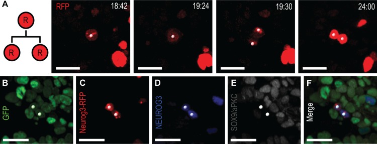
Fig 6. A small number of Neurog3-RFP cells divide into two NEUROG3+ cells.
(A) Still images from S8 Movie, demonstrating division of Neurog3-RFP cell (white spots) in RFP channel from a Pdx1 tTA/+;tetO-H2B-GFP;Neurog3-RFP explant. (B–F) Images of fixed explant with native GFP (B) and immunostained for Neurog3-RFP (C, staining for Myc-tag), NEUROG3 (D) and Sox9/aPKC (E). Both daughters are NEUROG3+/RFP+. Numbering denotes elapsed time in h:min (A). Scale bars, 20 μm.

