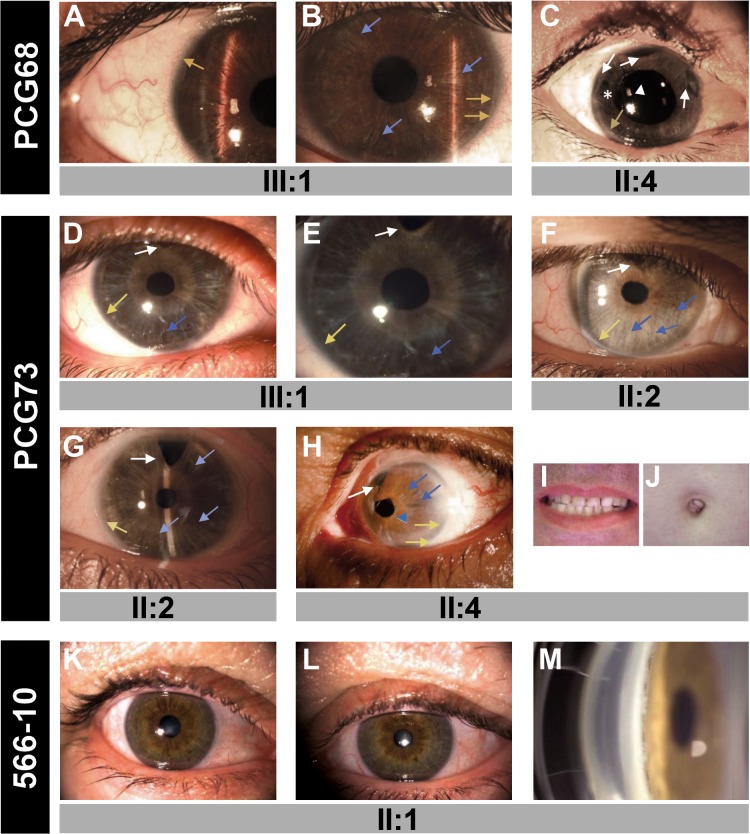
Fig 2. The phenotypes associated with the FOXC1 mutations identified in this study.
(A) Right and (B) left eye of subject PCG68 III:1. (C) Right eye of subject PCG68 II:4. (D) Right and (E) left eye of proband PCG73 (subject III:1). (F) Right and (G) left eye of subject III:1. (H) Axenfeld-Rieger anomaly in the left eye of subject PCG73 II:4. (I) Normal teeth and (J) umbilicus of this patient. (K) Right and (L) left eye, and (M) gonioscopy photography of subject 566–10 II:1. Asterisk: area of iris atrophy secondary to surgery. Blue arrows: areas of thin iris. Blue arrowhead: corectopia. Yellow arrows: posterior embrytoxon. White arrowhead: Ahmed valve tube in anterior chamber. White arrows: surgical iridectomies.

