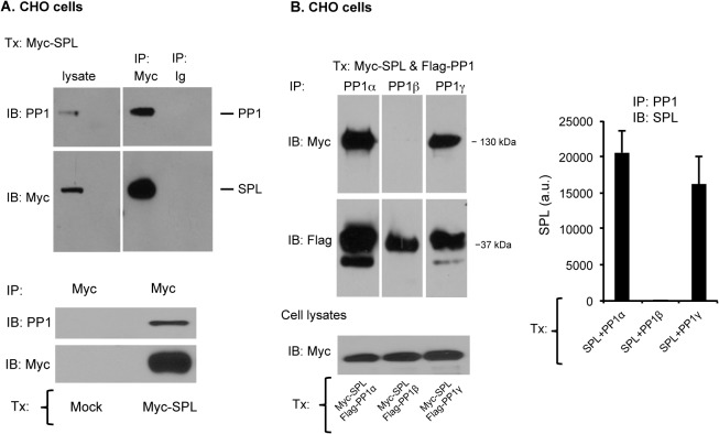
Fig 2. Formation of the SPL/PP1 complex in CHO cells.
(A) Top: Lysates were prepared from CHO cells transfected with a full length, Myc-tagged spinophilin (Myc-SPL). Proteins were precipitated with an anti-Myc antibody or nonimmune immunoglobulin (Ig) and then probed for PP1 and Myc-SPL. Bottom: Lysates were prepared from mock-transfected control CHO cells or cells transfected with a full length, Myc-tagged spinophilin (Myc-SPL). Proteins were precipitated with anti-Myc and then probed for PP1 and Myc-SPL. (B) Lysates were prepared from CHO cells transfected with full length Myc-SPL and either PP1α, β or γ. Proteins were precipitated with PP1 isoform-specific antibodies and then probed for Myc-SPL or Flag-PP1 (mean ± SEM, N = 3). Note that all the samples were run on the same gel and, as indicated by the vertical gaps, two marker lanes were excised.

