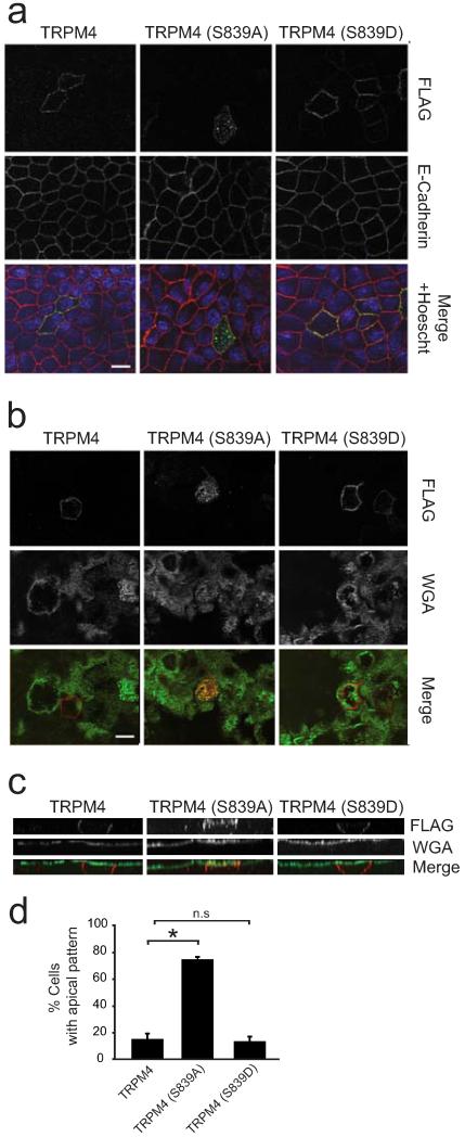
Fig. 4. Phosphorylation at the S839 residue is required for proper TRPM4 basolateral localization.
MDCK cells were grown as confluent polarized monolayers after transfection with TRPM4, TRPM4 S839A or TRPM4 S839D. The apical domain was labeled prior to fixation with WGA. After fixation, cells were immunostained with pAb anti-FLAG (green) and mAb anti-E-cadherin. Nuclei were stained with Hoechst (blue). Scale bar, 10 μm. a. Monolayer of MDCK cells focused at a section midway through the cells, the basolateral domain is labeled for E-cadherin (red). b. Monolayer of transfected MDCK cells focused at the apical domain, as labeled with WGA (red). c. Lateral view of monolayer showing the differences in localization between the different versions of TRPM4. d. Graph shows data on percentage of cells which exhibited apical localization of TRPM4 isoforms. * corresponds to significant differences (ANOVA test, post-Dunnett, p < 0.01) versus wild-type TRPM4. Data are from 3 independent experiments including n=559, 498, and 417 cells for TRPM4, TRPM4 S839A, and TRPM4 S839D, respectively.

