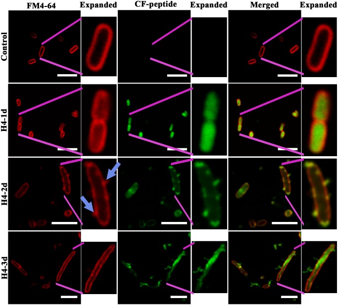
Fig 3. Confocal images of E. coli on treatment with HBD4 analogs.
Localization of caboxyfluorescein (CF) labelled peptides H4-1d, H4-2d and H4-3d in E. coli stained with inner-membrane dye FM4-64. Expanded images for bacteria are also shown adjacent to each panel indicated by pink lines. Blue arrows in FM4-64 expanded panel show membrane protrusion and lipid aggregation due to H4-2d. Scale bars represent 5 μm.

