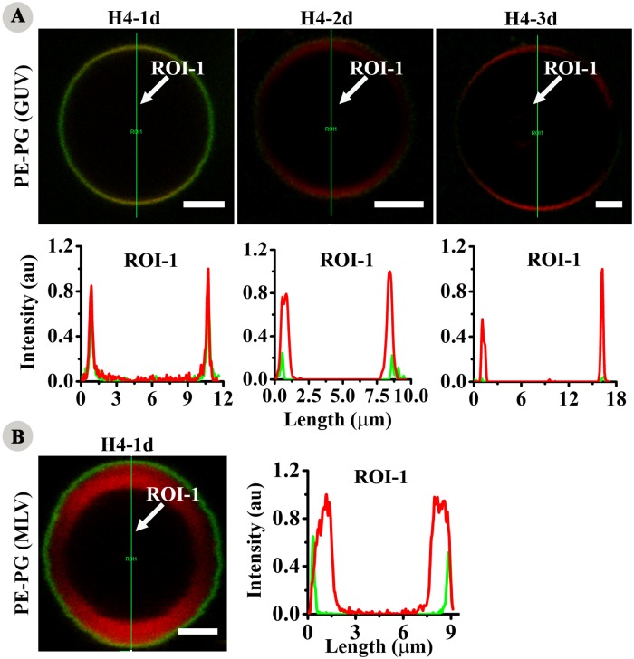
Fig 8. Interaction with lipid vesicles.
Localization of CF-labelled analogs with anionic giant unilamellar vesicles (GUVs) and multilamellar vesicles (MLVs) composed of lipids PE and PG in the molar ratio of 7:3 doped with Rh-PE. Green and red fluorescence represent CF-labelled peptide and rhodamine labelled vesicles respectively. Intensity vs length spectra show fluorescence intensity along the lines (ROI-1) drawn across the respective vesicles. (A) localization in GUV and (B) localization in MLV. Scale bars represent 2.5 μm.

