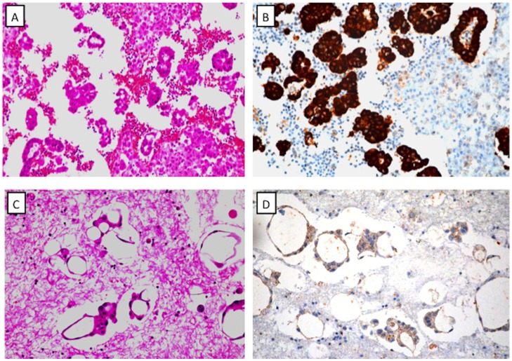
Fig 1. Representative examples of Ventana immunohistochemical staining of ALK.
A: H&E staining of a cell block shows lung carcinoma cells. B: Strong staining of ALK protein in a cell block slide (corresponding to sample A). C: H&E staining of a cell block shows lung carcinoma. D: Weak staining of ALK protein in a cell block (corresponding to sample C).

