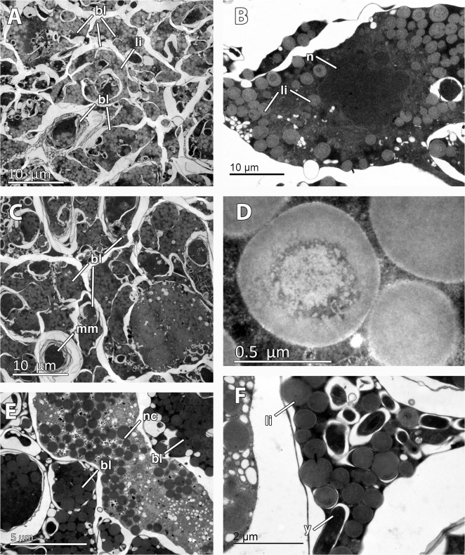
Fig 5. Embryonic features of Mycale acerata.
A. General view of inner blastomeres (bl) of the embryo, containing abundant lipid droplets (li). B. Detail of nucleated (n) blastomere showing lipid droplets (li). C. Multimembrane inclusions (mm) in the blastomeres (bl) of the embryo. D. Detail of lipid droplet. E. Embryonic cell similar to nurse cell (nc) among the embryo blastomeres (bl). F. Detail of rod-shape inclusions (y) in the blastomeres.

