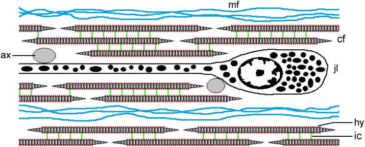
Fig 7. Diagrammatic representation of the CD of P. lividus.
This depicts part of a longitudinal section; not to scale. The CD consists of parallel discontinuous collagen fibrils (cf), which are connected by interfibrillar crossbridges containing chondroitin sulphate/dermatan sulphate proteoglycans (ic). Hyaluronic acid (hy) is present but of unknown function (its disposition on the surface of the collagen fibrils is conjectural). Bundles of beaded microfibrils (mf) occur between the collagen fibrils. The main cellular components are the somata and processes of electron-dense granule-containing juxtaligamental cells (jlc). Agranular cell processes, which may be cholinergic axons, are in close contact with the juxtaligamental cells and are shown as transverse sections (ax).

