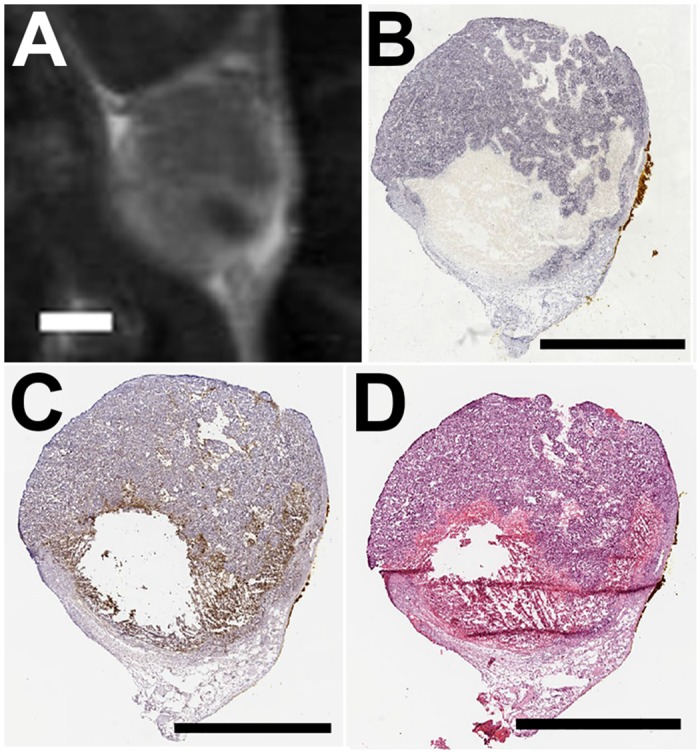
Fig 4. T1w contrast enhanced image at 48 hours post ablation and corresponding serial cryosections.

(A) Contrast enhanced T1w image of a tumor 48 hours following HIFU ablation. The under perfused region is visible as the dark spot in the lower right corner of the tumor. Serial sections were stained for (B) NADH diaphorse, (C) caspase 3, and (D) H&E. Scale bar represents 2 mm in each case. All panes present images of the same animal. Histological sections were acquired immediately following 48 hour MR imaging (A) and were registered with the image by using the skin interface as a reference point.
