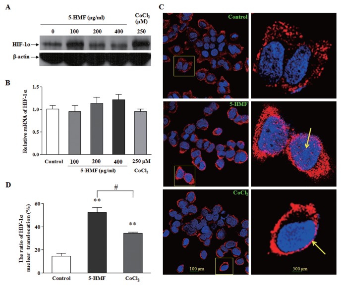
Figure 2.
The increased stability and nuclear translocation of HIF-1α by 5-HMF in PC12 cells. (A) Effects of 5-HMF on the levels of the HIF-1α protein. PC12 cells were exposed to the indicated concentrations of 5-HMF or CoCl2 for 1 h. The representative immunoblots are shown. (B) Effects of 5-HMF on the levels of HIF-1α mRNA. The HIF-1α mRNA levels were determined by qRT-PCR analysis after cells were treated for 1 h. The relative expressions were normalized to the control level. (C) Effects of 5-HMF on the nuclear translocation of HIF-1α. The distribution of the HIF-1α protein was analyzed with immunofluorescence staining after the cells were treated with 100 μg/mL 5-HMF for 1 h. The cells were labeled for HIF-1α (red) and counterstained with DAPI (blue), which was used to label nuclei. The arrowheads indicate the nuclear localization of HIF-1α, which was stained pink. The image at the right panel is the enlarged image in the frame at the left. (D) Statistical analysis of the ratio of HIF-1α nuclear translocation. The ratio was calculated in terms of the following formula: HIF-1α nuclear translocation (%) = (the number of cells with pink/the total number of cells with red) × 100. The results are expressed as the means ± SE from three independent experiments. **p < 0.01 compared with the control; #p < 0.05 for comparisons between the indicated groups.

