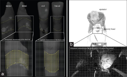
Figure 3.

(a) Illustration of the measurement of subgingival residual plaque of tooth 31 after working with a sonic scaler (AIR) for 120 s. The enlargements show the measurement with the standardized data masks with quadrangle arrays. (b) For demonstration purposes; the experimental setup in the darkened treatment room under inverse light conditions with the operator in 12 o’clock treatment position. The enlargement shows part of a photo taken under experimental conditions to measure the spatter distribution
