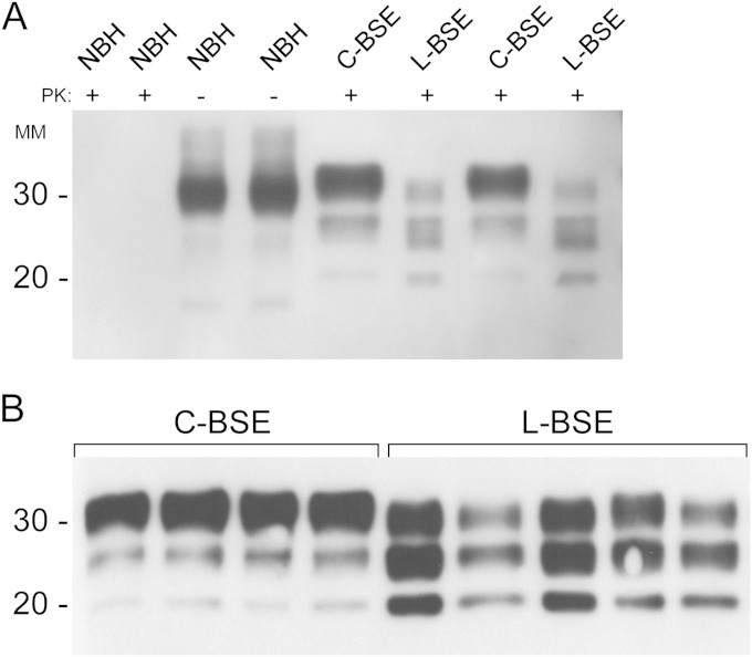
FIG 1.
Western blots of brain stem and frontal cortex samples from normal, C-BSE-infected, and L-BSE-infected cattle. (A) Two brain samples from normal cattle (NBH) were subjected to immunoblotting without proteinase K (PK) digestion (NBH −PK lanes). The same two brain samples from healthy cattle along with two brainstem specimens from C-BSE-positive and two frontal cortex samples from L-BSE-positive cattle were PK digested as described in Materials and Methods (NBH, C-BSE, and L-BSE +PK lanes). (B) Homogenized brainstem specimens from four C-BSE-positive and frontal cortex samples from five L-BSE-positive cattle were treated with proteinase K (PK) and subjected to immunoblotting using monoclonal antibody 6H4 (epitope within PrP residues 144 to 152). MM, molecular mass (in kilodaltons). The data are representative of multiple immunoblots of these specimens.

