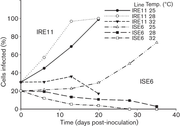
Fig. 1.

Growth of ISO7T in cell lines IRE11 and ISE6. Infected IRE11 cells were diluted 1 : 5 with uninfected cells and inoculated into replicated cultures. Infection was monitored by preparing cytocentrifuge slides from each culture at selected times for staining with Giemsa stain. Spread of infection was measured by determining the proportion of infected cells at each time point. The values at each time point are the mean of two replicated cultures.
