
Fig. 2.
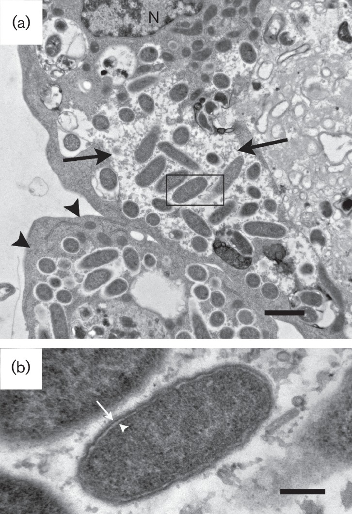
Transmission electron microscopy of IRE11 cells infected with ISO7T. (a) I. ricinus (IRE11) cell with ISO7T in cytoplasm (arrowheads) and in a vacuole (arrows). N, host cell nucleus. Bar, 1 µm. (b) Rickettsiae free in the cytoplasm showing the outer microcapsular layer, cell wall (arrow), electron-lucent periplasmic space and the periplasmic membrane (white arrowhead). Bar, 0.2 µm. The rectangle in (a) delineates the area shown in (b).