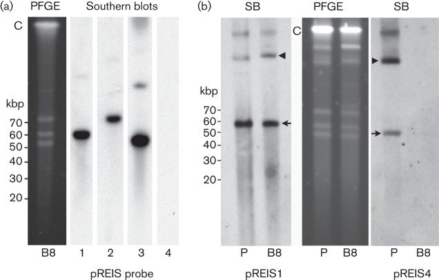
Fig. 3.
PFGE and Southern blot (SB) analysis of parental isolate ISO7T and clone B8. Cells were released by passing infected IRE11 cells through a 25 G needle. The suspension was filtered through a sterile 2 µm syringe filter and prepared for PFGE and Southern blot analysis (Burkhardt et al., 2011). (a) B8, PFGE and Southern blots probed with digoxigenin-labelled parA probes specific for plasmid pREIS1, -2, -3 or -4. Note absence of pREIS4. (b) Parental ISO7T (P) and clone B8 PFGE gel and Southern blots. Blots were probed with digoxigenin-labelled parA probes specific for plasmid pREIS1 or pREIS4. Note presence of pREIS4 in the parent (P). Arrows mark putative monomers of each plasmid and arrowheads indicate their conformational isomers. C, chromosomal DNA. Linear DNA marker positions are to the left of panels (a) and (b).

