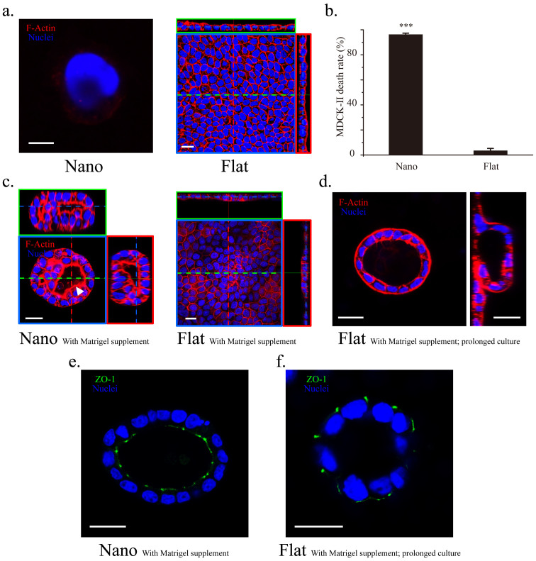
Figure 3. MDCK-II cells responded to nanotopography distinctly from Calu-3 cells.
(a) Co-staining of F-actin (red) and the nuclei (blue) showed that a single MDCK-II cell died after 1 d when cultured on the nanograss-patterned substrate (left, F-actin is almost undetectable); by contrast, MDCK-II cells formed a confluent monolayer on the flat substrate after 3 d in culture (right). (b) Death rate of adherent MDCK-II cells after incubation for 12 h on the nanograss-patterned and flat substrates (n = 3 independent experiments, each with 5 populations of cells; ***, P = 9.92E−5 compared to the flat substrate). (c) MDCK-II cells formed spheroids with lumens on the nanograss while maintained as monolayer on the flat substrates with 2% Matrigel supplement in the culture medium on Day 3. The lumen (white arrow head) is partially filled with fragmented nuclear material. (d) On Day 6, prolonged culture of MDCK-II cells on the flat substrates with Matrigel supplement generated lumens by buckling and wrapping of the cell sheet (left, top view; right, side view). (e, f) The lumens generated on the nanograss and the flat substrates featured a normal and a reversed polarity, respectively. Scale bars, (a): 5 μm (left) and 20 μm (right); 20 μm (c–f).

