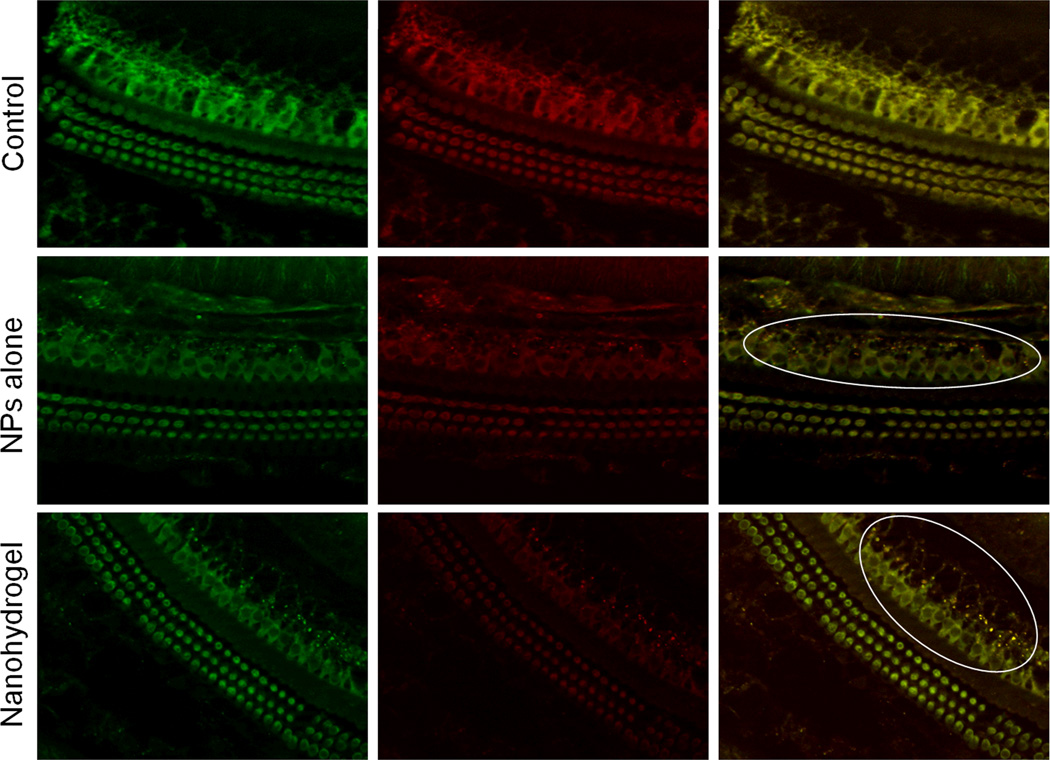
Figure 6. Microscopic evaluation of the cochlea following nanohydrogel application.
Twenty four hours after nanohydrogel was applied onto the round window niche in vivo, NPs were detected in inner ear cells as evaluated by the CF (Left - Green) and Rhodamine (Center - Red) fluorescent signal colocalization (Right - Yellow dots) (Right). The right ears treated with mock surgery served as controls. White ovals were drawn to highlight the NPs. (Original magnification 40×)

