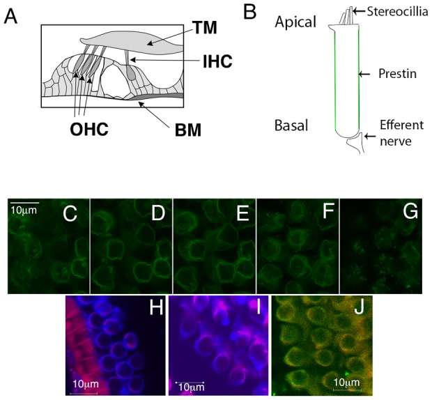
Fig. 1. Prestin in mouse outer hair cells is localized along the lateral wall of the cell along with β-catenin and Na/K ATPase.

Shown are cartoons of the organ of Corti (A) and its contained outer hair cells. TM, tectorial membrane; BM Basilar membrane; IHC, inner hair cell; OHC Outer hair cell. (B) A model of an outer hair cell in which prestin is shown lining its lateral wall (green). (C–G) The figure shows serial X-Y sections of mouse outer hair cells labeled with an anti-prestin antibody and then visualized using a Leica gated STED microscope. The sections start at the apical end (left) and end at the basal end (right). There is an uniform labeling of prestin along the lateral wall of the cell (middle three panels). Prestin labeling at the apical end of the cell tapers (C). Similarly, there is a patchy clustering of prestin at the basal pole of the cell (G). Mouse outer hair cells immunostained with antibodies to the basolateral markers β-catenin (H) and Na/K ATPase (I), and demonstrates the localization of these proteins (blue) along the lateral wall of the cell. The cells were counterstained with phalloidin Alexa 546 (red), which shows the presence of the sub cortical lattice of actin along the lateral wall of the cell. (J) The AP1µ1B subunit (green) is present in outer hair cells evidenced by antibody labeling of these cells. The figure shows co labeling of these cells with Na/K ATPase (red). Scale bar is 10 microns. These experiments were repeated five times.
