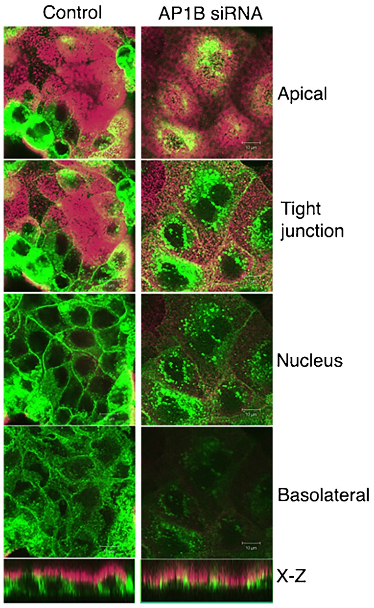
Fig. 8. Targeting of prestin YFP to the basolateral membrane requires AP1B (μ1B).

MDCK cells were electroporated with prestin YFP plasmid and siRNA to AP1B (μ1B). Cells were plated at confluent density and fixed after 30 hours. Cells were stained with antibodies to the apical marker GP130 and visualized by confocal microscopy. Shown are X-Y images along the z axis of transfected cells. The lowest panel shows the corresponding X-Z sections. Transfection of MDCK cells with siRNA to AP1B (μ1B) resulted in an apical localization of prestin YFP and near absence in targeting to the basolateral surface. In contrast, cells co-transfected with control siRNA (chicken KCNMB4) resulted in basolateral targeting of prestin YFP. The scale bar is 10 microns.
