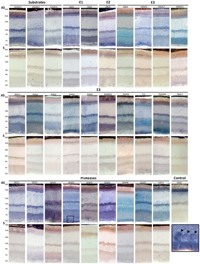
Fig. 3. In situ hybridization on murine retina cryosections of the genes encoding SUMO substrates and SUMO E1, E2, and E3 ligases, and proteases.
Representative images obtained after the in situ hybridization of Antisense (AS) and Sense (S) digoxigenin-labelled riboprobes, stained for the same period of time per each gene. Antisense riboprobes reflect the pattern of gene expression, whereas the sense probes are the corresponding negative controls. The antisense Rhodopsin probe, which strongly labels the inner photoreceptor segment, was used as a positive control for the assay. RPE, Retinal Pigment epithelium; PhR, Photoreceptor cell layer; ONL, outer nuclear layer; OPL, Outer plexiform layer; INL, Inner nuclear layer, IPL, inner plexiform layer; GCL, ganglion cell layer. The boxed region at the bottom right is an amplification of the Senp2 in situ hybridization at the GCL level. The black arrowheads indicate nuclear/perinuclear mRNA localization.

