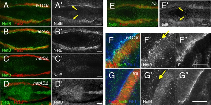
Fig. 1. Netrin internalisation on the basal side of midgut cells requires Fra.

Stage 13 embryos immunostained with Fas3 (red A–G) to identify the VM, NetB (green A–G; grey A′–G′) and Fil-1 (blue, F–G; grey F″–G″). (A) NetB is expressed in the visceral mesoderm (A′, brackets) and is basally enriched in the midgut cells (arrows) (n>50 embryos, Note: unless otherwise stated embryos depicted in figures exhibit phenotypes representative of all observed embryos). Anterior is to the left for all embryos (and throughout this paper). (B) netAΔ embryo. NetB expression appears normal (n = 18 embryos). (C) netBΔ embryo. No midgut-specific expression in observed (n = 4). (D) netABΔ embryo. No tissue-specific expression is observed (n = 6). (E) fra3/Df(2R)BSC880 embryo. NetB expression is seen in the VM, but the line of enrichment towards the basal end of midgut cells is lost (arrows) (n = 6). (F–G) High-resolution images of NetB localisation. NetB is expressed in the VM and is enriched within the midgut cells towards their basal end nearest the VM (F′, arrow). (G) fra3/Df(2R)BSC880 embryo. NetB is lost from the basal side of the midgut cells (G′, arrow). Scale bars, 20 µm.
