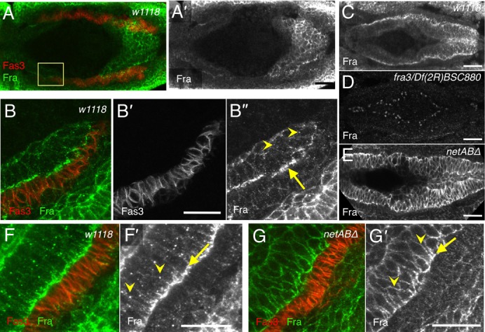
Fig. 2. Fra basal polarisation and internal puncta are dependent upon Netrins.

Stage 12 (A,B) and stage 13 (C–G) embryos stained with Fas3 (red; grey B′) and Fra (green; grey A′,B″,C–E,F′,G′). (A) Fra is expressed in the migrating midgut primordia (n = 12). (B) High-resolution image of the boxed region indicated in A. Fra expression is enhanced on the basal side of the midgut cells (B″, arrow), and in some intracellular punctae (B″, arrowheads). (C) Fra is strongly, basally polarised by stage 13, and is present in intracellular punctae (n = 13). (D) No Fra could be detected fra3/Df(2R)BSC880 embryo (n = 5). (E) netABΔ embryo. Fra expression becomes increased in the lateral membranes, and the intracellular punctate expression is lost (n = 21). (F) In a w1118 embryo Fra is enriched within midgut cells towards their basal surface (F′, arrow) and localises to intracellular punctae (F′, arrowheads). (G) In a netABΔ embryo basal polarisation is less pronounced (arrow), levels on lateral membranes are increased (arrowheads) and punctae are lost. Scale bars, 20 µm.
