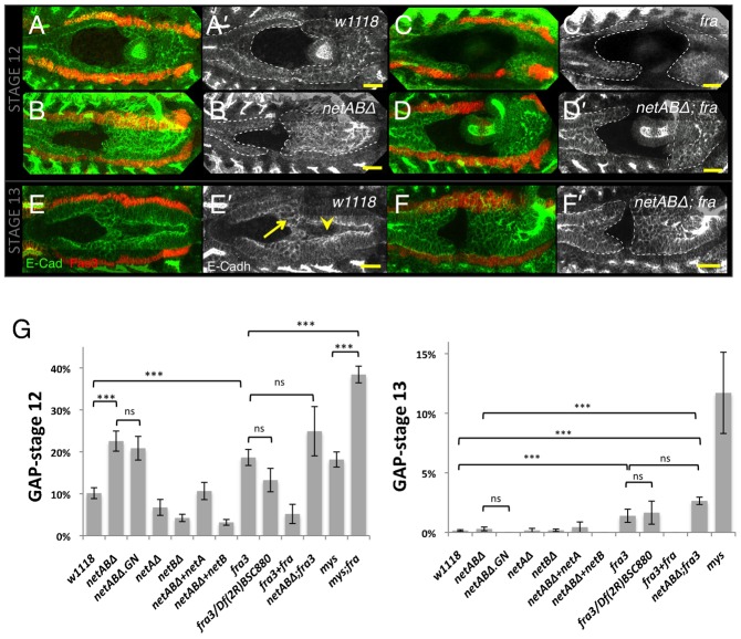
Fig. 4. Embryonic midgut migration is delayed in netrin and fra mutants.
(A–F) Stage 12 (A–D) and stage 13 (E–F) embryos immunostained for Fas3 (red) to identify the VM, and for E-Cadherin (green; grey A′–F′) to identify the midgut. Dotted line depicts extent of midgut. (A) w1118 control embryo with the midgut primordia just meeting. (B) In a netABΔ embryo migration is delayed. (C) fra3/Df(2R)BSC880 mutant embryo showing a greater migration delay. (D) Combined loss of netrins and fra enhanced the delay phenotype. (E) Stage 13 w1118 control embryo. The epithelium has formed, and only ICPs (arrow) and AMPs (arrowhead) are yet to incorporate into the epithelium. (F) netABΔ;fra3 mutant. A gap between the primordia is still evident. Note: The VM was well formed and continuous in all of these genotypes. Any apparent breaks are due to the VM being outside the focal planes shown. (G) Quantification of migration delay in stage 12 and stage 13 embryos. For p-values and n-values see Table 1. Scale bars, 20 µm. *** = p<0.001, ns = p>0.05.

