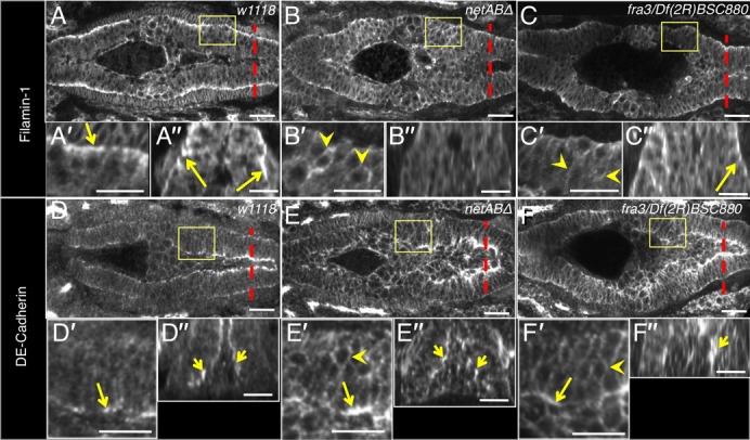
Fig. 6. Netrins and Fra are required for the midgut MET.

Stage 13 embryos showing disruption of epithelium formation. Boxed regions in (A–F) are magnified in (A′–F′), and cross-sections taken at the dotted lines are shown in (A″–F″). (A) w1118 control embryo. Filamin-1 is basally polarised (arrows; A′–A″) (n = 27). (B) netABΔ embryo. Basal polarisation of Filamin-1 is lacking (B′–B″). Instead it is distributed around the entire cell membranes (arrowheads) (n = 28). (C) fra3/Df(2R)BSC880 embryo. Basal polarisation is reduced though not absent (C″, arrow) and expression is increased around the entire cell membranes (arrowheads) (n = 14*). (D) w1118 embryo. E-Cadherin is apically polarised in the midgut cells (arrows) (n = 10). (E,F) In netABΔ embryo (n = 9) and fra3/Df(2R)BSC880 embryos (n = 10) E-Cadherin apical localisation is reduced but still apparent (arrows) and shows increased expression around the entire cell membranes (arrowheads). * for fra mutants, n-values are pooled from fra3/Df(2R)BSC880 and fra3/fra3 genotypes which exhibited the same phenotype. Scale bars, 20 µm.
