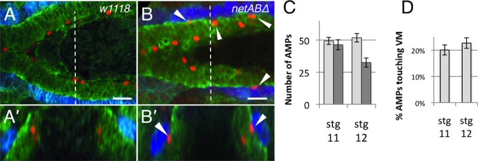
Fig. 7. Adult midgut precursor cells are mislocalised in netABΔ mutant embryos.

(A,B) Stage 12 embryos immunostained with Fas3 (blue) to mark the VM, Filamin-1 (green) to mark the midgut and Asense (red) to mark the Adult Midgut Precursor (AMP) cells. Images show only the anterior midgut. (A′–B′) represent cross sections taken at the dotted line in (A,B). (A) The AMPs in w1118 embryos are located on the apical surface of the developing midgut epithelium. None come into contact with the VM. (B) In netABΔ embryos, some AMPs are found in contact with the VM (arrowheads). (C) Quantification of AMP numbers in w1118 (light grey) (n = 3, n = 5, at stg 11, 12 resp.) and netABΔ embryos (n = 3, n = 4, at stage 11, 12 resp.). (D) Proportion of AMPs in contact with the VM in netABΔ embryos. Scale bars, 20 µm.
