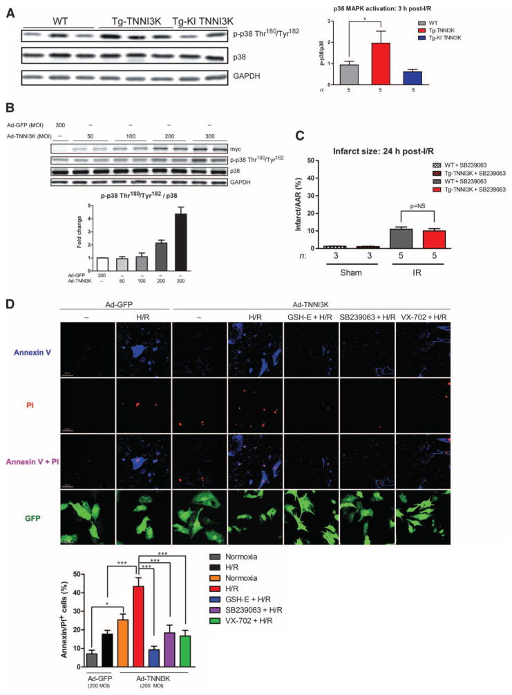
Fig. 4. TNNI3K promotes I/R injury and cardiomyocyte death through increased p38 MAPK activation.
(A) Levels of phosphorylated and total p38 MAPK in lysates from the ischemic LV of Tg-TNNI3K or Tg-KI TNNI3K mice, 3 hours after I/R. GAPDH served as a loading control for total protein. Quantification from n = 5 animals per group is shown. (B) Levels of phosphorylated and total p38 MAPK in lysates from NRVMs infected with increasing MOI of adenoviruses expressing either N-terminal myc-tagged TNNI3K or GFP. Levels of myc epitope served as an indicator of TNNI3K overexpression, and GAPDH was a loading control. Quantification from n = 3 biological replicates is shown below the immunoblots. (C) Infarct sizes 24 hours after I/R in Tg-TNNI3K or WT mice treated with the p38 inhibitor SB239063 1 hour before I/R. (D) Representative confocal micrographs from n = 3 biological replicates of intact NRVMs labeled with propidium iodide (PI) and annexin V, after H/R. Cells were infected with Ad-TNNI3K or Ad-GFP control 24 hours before H/R. Cells were treated with glutathione ester (GSH-E) or SB239063 or VX-702 1 hour before H/R. Scale bars, 20 μm. Quantification of cell death (as percent of PI+/annexin V+ cells) is shown. For all graphs, data are means ± SEM. *P < 0.05, ***P < 0.001 as determined by one-way ANOVA followed by Tukey’s post hoc test.

