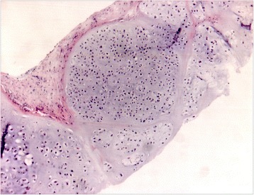
Figure 2.

Tru-cut biopsy of the pelvic, primary tumour, performed in December 2009. Histopathological examination (HE x5, inset x10): fibrous tissue with nests of cartilaginous proliferation with hypercellularity and variation in cellular size and shape, in a focally myxoid matrix. Final diagnosis was G2 peripheral conventional chondrosarcoma. Radiologic features were not consistent with the presence of dedifferentiated areas thus supporting the final diagnosis of a conventional chondrosarcoma.
