Figure 3.

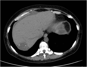
CT scan without contrast of the liver at the time of the first hepatic progression, showing a single metastasis, characterised by pronounced hypodensity and calcification islets (axial plane, abdomen window).

CT scan without contrast of the liver at the time of the first hepatic progression, showing a single metastasis, characterised by pronounced hypodensity and calcification islets (axial plane, abdomen window).