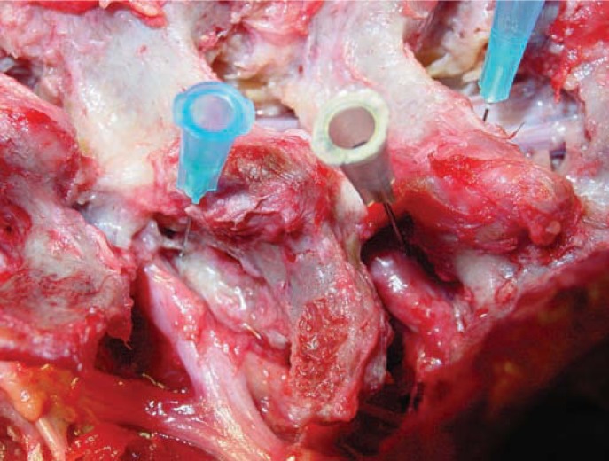
Figure 1.
Foraminal view of cadaver dissection at L4–5 and L5–S1. Note the tiny furcal nerve branch speared by the blue hubbed needle that branches from exiting L4 nerve at L4–L5. The nerve seen in Figure 5b would be what a small furcal branch looks like, but in this instance, excisional biopsy confirmed by pathology slide revealed the nerve to be an autonomic nerve because of the ganglia seen on the slide. These furcal nerves in the “hidden zone” of MacNab vary in size and can cause postoperative dysesthesia. When the nerve is equivalent in size to the spinal nerve, it can be interpreted as a conjoined nerve if it cannot be traced back to its origin.

