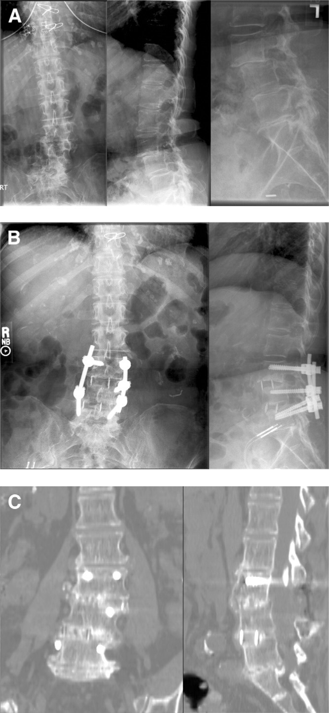
Fig. 2.
Preoperative (top) radiographs, immediate postoperative (middle) radiographs, and 24-month (bottom) CT scans of a 68-year-old female anteriolateral fusion patient treated for degenerative spondylolisthesis and scoliosis. Note increase in disk height, correction of coronal and sagittal alignment, and maintenance of corrections as well as bridging bone in 24-month images.

