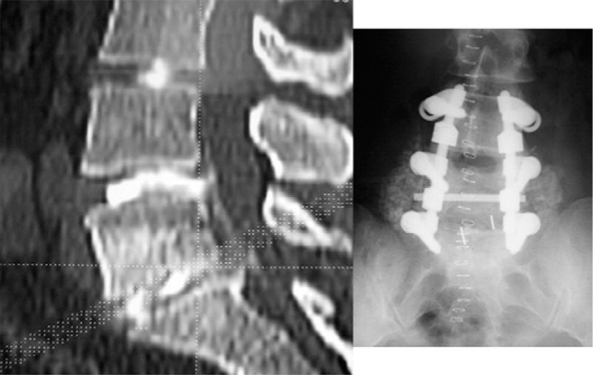
Fig. 4.
Left, Discogram showing pathologic morphology at both the L5-S1 and the L4-5 levels. A non-painful level with normal architecture was mandatory at the L3-4 cephalad segment. Right, Computed tomography discogram with pathologic morphology at both L4-5 and L5-S1. Both of these levels are fully concordant; the L3-4 level presents normal morphology and a negative discogram. This patient underwent an L4-5 fusion with dynamic instrumentation at L4-5.

