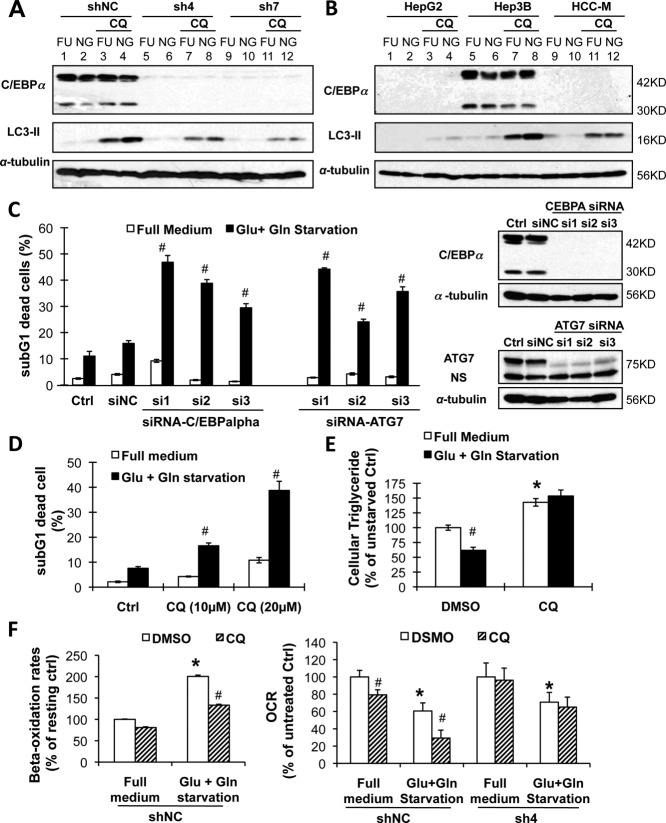
Figure 5.
Autophagy was essential for C/EBPα-mediated lipid catabolism and protection against starvation. (A) Cells expressing C/EBPα (shNC) and C/EBPα-silenced stable cells (sh4 and sh7) were treated with or without 25 µM of the lysosomal inhibitor CQ in glucose- and glutamine-free medium for 3 hours. Flux of LC3-II was determined by western blotting. (B) Cells expressing C/EBPα (Hep3B) and C/EBPα-deficient HepG2 and HCC-M cells were treated as in A. (C) The Hep3B cells were silenced of ATG7 or C/EBPα by specific siRNAs for 2 days before starvation treatment (siNC for nonspecific siRNA) for 3 days. Dead cells were determined by sub-G1 assay (left panel) and the proteins by western blotting (right panel). (D,E) The Hep3B cells were starved with or without CQ for 2 days. Dead cells were determined (D), and the intracellular triglyceride level was calculated as in Fig. 3B. (F) The shNC and sh4 cells were starved with or without CQ (100 µM). Fatty acid beta-oxidation rates were determined (left panel), and oxygen consumption rates were separately determined by mitochondrial stress assay (n = 4, right panel). Abbreviations: FU, full medium; NG, glucose- and glutamine-free medium; NS, nonspecific staining; DMSO, dimethyl sulfoxide.

