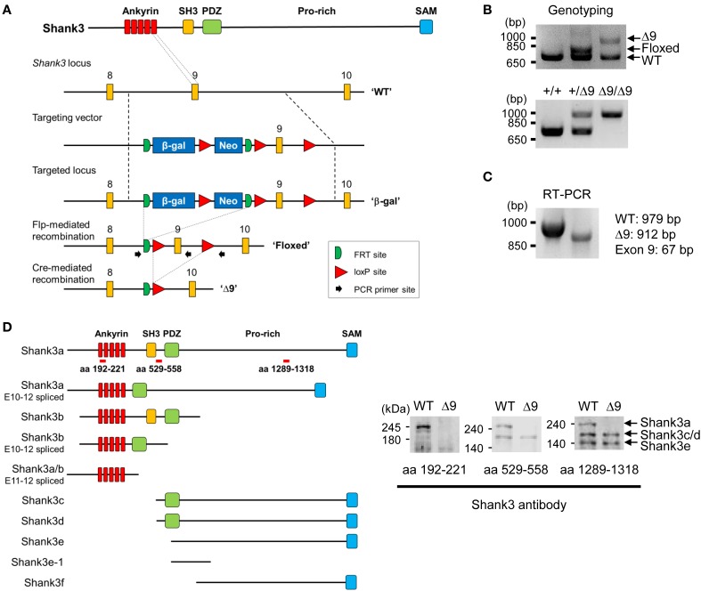
Figure 1.
Generation and characterization of Shank3Δ9 mice. (A) Targeting the Shank3 locus in mice and removal of exon 9 by Cre-mediated recombination. Ankyrin, ankyrin repeats; SH3, src homology 3 domain; PDZ, PSD-95, Dlg, ZO-1 domain; Pro-rich, proline-rich region; SAM, sterile alpha motif; β-gal, β-galactosidase; WT, wild-type. (B) Genotyping PCR of the targeted locus. Δ9, Shank3Δ9. (C) RT-PCR of WT and Shank3Δ9 mouse brains showing the removal of exon 9 at the mRNA level. (D) Immunoblotting of Shank3 splice variants in WT and Shank3Δ9 brains by three different antibodies. Targeted regions of the antibodies are indicated by red bars on the recently reported 10 Shank3 splice variants (Wang et al., 2014b). Note that the longest isoform (Shank3a band) is undetectable in the Shank3Δ9 brain.

