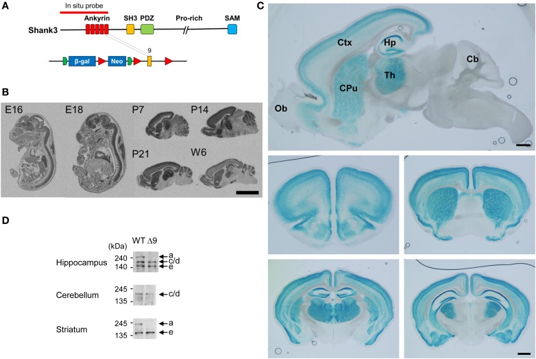
Figure 2.
Expression patterns of ankyrin repeat-containing variants of Shank3 mRNAs and proteins. (A) Locations of the in situ hybridization probe and β-galactosidase insertion. (B) Distribution patterns of ankyrin repeat-containing Shank3 mRNA variants in mouse embryonic and postnatal brain (sagittal) sections, as revealed by in situ hybridization. Scale bar, 5 mm. E, embryonic; P, postnatal day; W6, postnatal week 6. (C) Ankyrin repeat-containing Shank3 protein variants, as revealed by X-gal staining of sagittal (top) and coronal (middle and bottom) Shank3+/β-gal brain sections (6–7 weeks). Ob, olfactory bulb; Ctx, cortex; Hp, hippocampus; Th, thalamus; CPu, striatum; Cb, cerebellum. Scale bar, 1 mm. (D) Differential expression of Shank3 protein variants in different brain regions, as revealed by immunoblot analysis of WT and Shank3Δ9 brain lysates (3–6 months) with the Shank3 antibody (aa 1289–1318).

