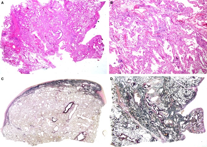
Figure 1.
A, A biopsy specimen from right S5 taken from a patient aged 69 years. Alveolar septa were diffusely thickened, with preservation of lung structure [haematoxylin and eosin (H&E)]. B, Alveolar septa were widened with septal fibrosis and a mild degree of inflammatory infiltration (H&E). C, An autopsy specimen taken from the right middle lobe, showing a fibroelastotic band just beneath the fibrously thickened pleura [elastica van Gieson (EVG)]. D, EVG staining of right S5 at biopsy. Lung parenchyma was rich in elastic fibres, with localized aggregates of elastic fibres and alveolar septal elastosis.

