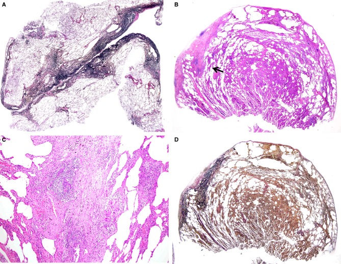
Figure 4.
A, A biopsy specimen from the superior segment of the left upper lobe taken from a patient aged 61 years, showing a typical feature of pleuroparenchymal fibroelastosis [elastica van Gieson (EVG)]. B, A biopsy specimen from right S2 taken at the age of 49 years, showing cellular interstitial pneumonia with localized areas of subpleural fibrosis [haematoxylin and eosin (H&E)]. C, Higher magnification of (B) (indicated by a black arrow), showing a granuloma with giant cells adjacent to the subpleural fibrosis (H&E). D, EVG staining of the sample shown in (B), revealing subpleural fibrosis consisting of fibroelastosis.

